Item Code: SU00103 Ultrasound Training Station
Brand: SimUltra

System Function
1.1. The virtual ultrasound training and assessment system has 3D anatomical structures and echocardiograms of various organs, combined with transthoracic (TTE), transesophageal (TEE), transvaginal, and linear array probe simulation ultrasound probes to simulate real ultrasound inspection scenes;
1.2. The analog ultrasound probe has a positioning sensor. The ultrasound probe can present the ultrasound image in real time when scanning the simulated person, and the ultrasound image can be changed synchronously by moving the angle of the ultrasound probe;
1.3. The system has multiple standard ultrasound sections, such as trans esophageal ultrasound section and transthoracic ultrasound section;
1.4. The simulated ultrasound probe can be used arbitrarily without restarting the device in actual training, including operations on normal and pathological hearts;
1.5. There are two scanning methods of virtual mouse control and analog probe control for ultrasound examination training and demonstration;
1.6. Support one-click freeze display of 3D anatomical views and corresponding ultrasound images, which is convenient for teaching;
1.7. The operation of the ultrasound simulator is divided into training and assessment modules, and each training module has teaching instructions;
1.8. The sharpness and contrast of ultrasound images can be set;
1.9. It has the function of measuring the distance, area and volume of organs;
1.10. The software has an ultrasound report template. Students can create an interactive ultrasound report, fill in patient information, measurement data, cardiac contractility data, description data, and save, upload and export;
1.11, has the function of saving screenshots of ultrasound images, and is stored in the system to support export use.
1.12, users can be created, and the training and assessment data of the trainees can be counted.
1.13. Use real medical couplant for ultrasound examination.
1.14, with 3D anatomical diagram virtual prompts, step-by-step video guidance.
1.15. The depth, angle and focal depth of the sensor can be changed in the system.
1.16. Ultrasound mode: including 3 ultrasound modes, cardiac ultrasound, color Doppler ultrasound and electrocardiogram.
Hardware Configuration
2.1. Model 1unit
2.2. High-profile computer host (with system) 1unit
2.3. LCD monitor 1 unit
2.4. Simulated ultrasound probes 3units
Gastroscope & ERCP Training Model
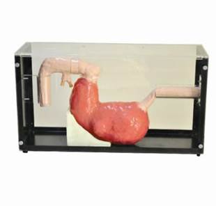
Features:
- Simulate the upper body of an adult. The form is lifelike and feels like real.
- It has obvious anatomic landmarks like nasal cavity, windpipe and bronchus. And it is elastic with high flexibility, which is convenient to localize.
- The model is applicable to operation training of trachea cannula of laryngoscopy through mouth, bronchofiberscopy and rigid tracheo-bronchoscopy.
- Head could lean back and do side-to-side movement, which is convenient for positions needed when operating.
- When conducting rigid tracheo-bronchoscopy and laryngoscopy, humming alarm rings when wrong operation of overexertion on teeth.
- Put stethoscope on two lungs to auscultate when conducting trachea cannula and bronchofiberscopy. Use rubber pressurized ball to simulate breath sounds to determine the position of bronchoscope catheter.
Price: xx zł
Vocal Cord Polyp Examination Model
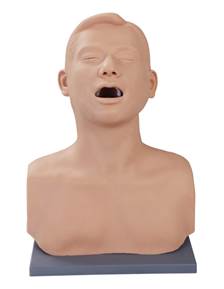
Features:
- Model for adult upper body, standard check position, open mouth.
- Vocal cord polyp examination.
Price: xx zł
Vocal Nodules Examination Model

Features:
- Model for adult upper body, standard check position, open mouth.
- Vocal nodules can be checked for operation.
Price: xx zł
Vocal Cord Tumor Examination Model

Features:
- Model for adult upper body, standard check position, open mouth.
- Vocal cord tumor operation
Price: xx zł
Electronic Throat Examination Model
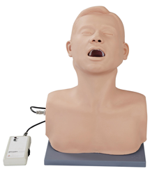
Features:
- Model for adult upper body, standard test position with mouth open.
- Soft tongue, can pull it out for facilitate inspection.
- Oral epiglottis can be seen 2 cm size of the cancer module, can use the spatula to view the lesion location and traits
- You can use the spatula to view the lesion location and traits
- When the operation is correct, external electronic box can prompt.
Price: xx zł
Ocular Inspection Model of Retinopathy

Features:
Replacement of different eyeground diseases, slide simulates common clinical oculopathy, including:
- Normal retina
- Age-related retina macular degeneration/colloid Bodies
- Central retinal vein occlusion
- Hypertensive retinopathy
- Papilledema
- Excavation of optic disc
- Optic atrophy
- Mild background diabetic retinopathy
- Background (pure) diabetic retinopathy
- Preproliferative diabetic retinopathy
- Preproliferative diabetic retinopathy 2
- Hypertrophic diabetic retinopathy
- Diabetic retinopathy
Basic configuration:
- Realistic affection, easy to replace and operate;
- In favor of repeated contrast observation;
- Provides 13 kinds of retinopathy accessories;
Price: xx zł
Electronic Retina Inspection Model
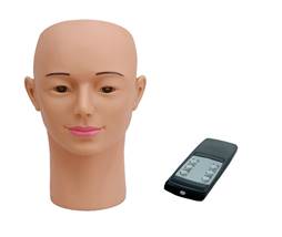
Features:
- Replica of an adult head and neck, with delicate five sense organs and standard position for fundus examination.
- Supports retina and various common fundus lesions examination
- Simulates clinical eyes by auto-changing slides of different fundus lesions
Price: xx zł
Electronic Ear Inspection Simulator
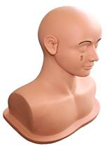
Features:
- Accurate anatomical structure such as auricle, external auditory canal, eardrum, etc.
- Inspect ear lesions with otoscope.
- Allow to practice in cleaning the earwax.
- Comes with over 14 negative films of ear lesions, no need to replace all the ear module, easy to operate.
Price: xx zł
Ear Inspection Simulator
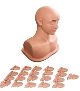
Features:
- Simulation of right ear, standard intra-aural examination site
- Accurate anatomical structure of auricle, external auditory canal, tympanic membrane;
- Ear lesion examination via otoscope;
- Provide 24 kinds of ear lesion components for convenient replacing;
- Including:
- Normal tympanic membrane
- Retracted tympanic membrane
- Small tympanic membrane perforation
- Whole tympanic membrane perforation
- Traumatic perforation of tympanic membrane
- Dry central perforation of the posterior
- Myringotomy with insertion of tube
- Bullous myringitis
- Herpes blisters on the tympanic membrane
- Eardrum Tympanosclerosis
- Tympanosclerosis crescentic sclerosis plaques
- Serous otitis media effusion
- Congestive early acute otitis media
- Acute otitis media
- Purulent otitis media
- Chronic suppurative otitis media
- Pearl-tumor
- Foreign body in ear
- Ear washing and drops
- Earwax clean-up operation practice
- Ear lesions component easy to replace.
Price: xx zł
Ear Diagnostic Simulator
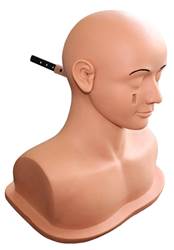
Features:
Standard body position of the ear, push/pull lesion to select lesions. Totally 12 kinds of normal or diseased ear drum:
- Acute otitis media
- Acute otitis media without obvious signs
- Abnormal secretion of middle ear
- Tympanosclerosis
- Myringotomy
- Ear wax (a little big)
- Acute middle ear infection
- Otitis media (case 1)
- Otitis media (case 2)
- Effusion of posterior ear drum
- Perforation of ear drum
- Normal tympanic membrane
Price: xx zł
Ear Irrigation Simulator

Features:
Practice irrigation of auditory canal to avoid the risk of direct irrigation for patient.
Price: xx zł
Debridement Suture Head Training Model
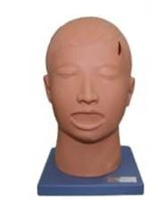
Features:
- Complete head model, realistic shape, soft skin.
- Skin layer clear, precise structure, the operation feel real, can be repeated suture practice.
- The model can touch the skull fracture, bilateral transverse maxillary fracture, I & III, nasal fracture, bilateral mandibular fracture, C-6 vertebral fracture, hemotympanum, deviated trachea, pupil teaching demostration
- The model provides a surgical incision for teaching.
Price: xx zł
Vasculature Operation Model

Features:
- Provide vein and artery blood vessels of different inner diameters to know different suture distance, tension/technique among main artery, vein and arteriole.
- High emulation and strongly elastic vessels
- Equipped with vessel fixator, easy to operate.
- Supports repeated practice.
Price: xx zł
Wound Hemostasis & Suture Module

Features:
- Comes with realistic skin tissue tension
- Six holes bleeding at the same time to simulate emergency ligation
- Supports repeated practice
Price: xx zł
Tenorrhaphy Model
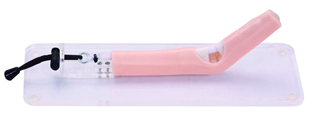
Features:
- Layered flexor tendon, can buckling any angle.
- Made from polymer material, environmental protection and no pollution, high-emulation.
- Equip skid proof pedestal.
- Support layered suture flexor tendon, tendon edge repaired and hypodermic needle tendon suture, etc.
Price: xx zł


